Why does a bruise turn purple, then green, then yellow?
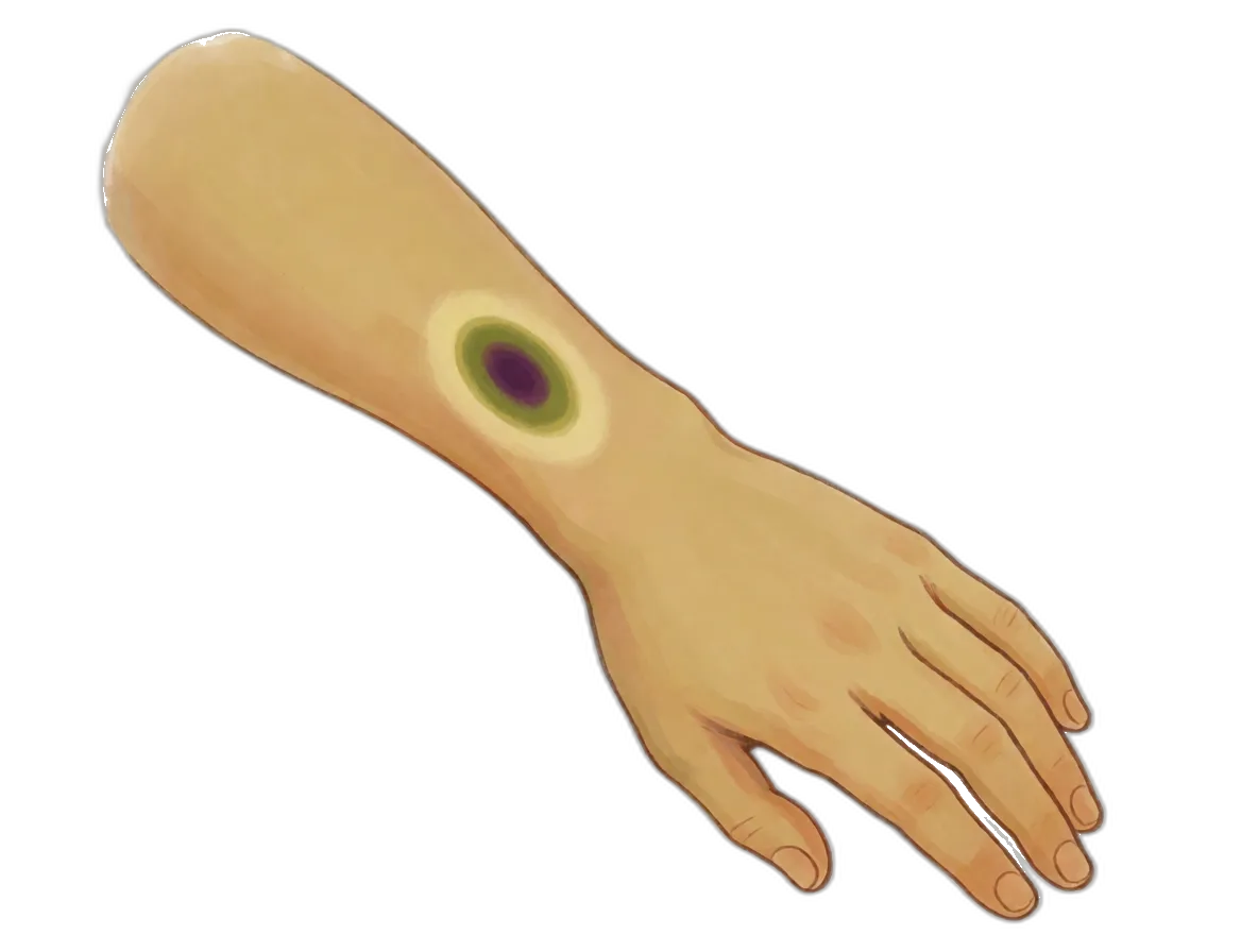
You catch your shin on the corner of a table. Nothing bleeds, nothing breaks, and a minute later you have forgotten it. Two days on, a dark purple oval is sitting exactly where you hit. By the weekend it has gone green, then a sickly yellow, then nothing. Every bruise you will ever have runs those colors in that order, so who chose them?
Nothing in that mark is your skin. It is blood, trapped somewhere it was never meant to be, and your body is taking it apart where it lies. Each color is one more piece coming off.
Blood with nowhere to go
Blood never sloshes around loose inside you. It travels in closed tubes, and the narrowest are the capillaries. Along them squeeze your red blood cells, flat discs crammed with the pigment that ferries your oxygen.
A red blood cell is wider than the narrowest capillary, so it folds up like a taco to get through.
Fine tubes have thin walls. Crack your shin on a table leg and your skin survives comfortably; the capillaries underneath do not. Their walls tear, and blood escapes into the surrounding tissue.
That blood is trapped. It cannot climb back into the tubes it fell out of, and it cannot leave through unbroken skin. It settles into a shallow puddle about 1–2 mm(1/16 in) down, and a bruise the size of a plum holds under 5 mL(1 tsp) of blood.
That teaspoon is deoxygenated. It handed its oxygen over hours ago, so it is darker than the blood circulating in your fingertip, nearer the color of a cherry. Which leaves one thing unexplained. Cherry red is still red, and the mark on your shin is purple.
Why red blood under skin looks purple
Skin is not a window. Light landing on your arm penetrates a short way, and scattering among the layers sends some of it back out. How deep it got depends on its color: red light travels furthest, blue scatters straight back out of the epidermis, the thin dead layer on the outside.
Over ordinary skin both kinds return, and the mixture is your normal arm color. Over a fresh bruise the red light reaches the puddle and must cross it twice, losing a great deal to absorption. The blue gave up long before it got near the blood.
Light leaving a bruise is therefore short of red, while the skin around it is not. Your eyes report the comparison, and skin short of red reads as purple.
Try it. Poke a hole in a scrap of paper, lay it over your wrist, and look at a blue line through it. Most of the blue drains away.
So the first color owes nothing to chemistry; part of it is your own eyes comparing one patch with another. Nothing in that puddle has altered yet. Give it a day, and something will.
Cleaners that eat the evidence
Spilled blood cannot be pumped anywhere. Your body sends cells that can crawl instead.
They are macrophages, from the Greek for big eaters, and eating is precisely the job. A macrophage is a shapeless white blood cell, several times wider than the discs it eats. It squeezes between other cells, flows around anything dead or out of place, and seals it inside itself.
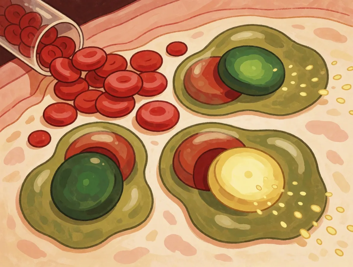
Within a day of the knock they arrive in thousands and swallow the spilled discs whole. Nothing is shipped elsewhere for processing: the eaters dismantle the mess where it lies, which is why the colors appear exactly where you banged yourself.
Clearing a decent bruise occupies them for the better part of two weeks. They begin at the edges, where the puddle is shallowest, and gradually work inward. A bruise a few days old is often purple in the middle with a green or yellow rim.
So the spill is taken apart inside cells, a mouthful at a time. The colors begin the instant it goes in, because of what waits inside the eater.
The red is a ring with iron at its center
Packed inside every red blood cell is hemoglobin, the pigment that collects oxygen in your lungs and delivers it everywhere else. Hemoglobin is a molecule: a fixed group of atoms, assembled identically every time.
Most of hemoglobin has no color whatsoever. The red is concentrated in four small flat rings buried inside it, and each of those rings is a heme. At the center of every ring sits a single iron atom, and that iron is the hook your oxygen clips onto.
Pigment molecules get their color from their shape. The arrangement decides which colors of light it absorbs and which it reflects, and this ring absorbs almost everything except red. Red escapes, so red is what you see.
So the redness of your blood is one small ring, repeated over and over inside every disc. Break the ring, and the red has to go somewhere.
Cut the ring open and it turns green
Inside the macrophage a tool goes to work on that ring. It is an enzyme: something a cell builds to make one exact change happen, over and over, and nothing else. This one is heme oxygenase — the cutter.
The cutter grips the ring at one spot and severs it. The closed loop springs open into a chain, and the iron drops out of the middle. Your body never discards that iron: it returns roughly 20 milligrams a day to circulation, which is why you need so remarkably little from your dinner.
Nearly all your iron is iron you have used before.
An open chain is a different shape from a closed ring, so it absorbs different colors. It absorbs red now and lets green through, the exact opposite of an hour earlier. That green pigment is biliverdin.
A second enzyme takes over almost immediately: biliverdin reductase, the finisher. It hooks two hydrogens onto the middle of the chain. That tiny alteration shifts the color from green to yellow, and the yellow pigment is bilirubin.
So one pigment goes in red, comes out green, and finishes yellow. The order can never swap, because the finisher has nothing to work on until the cutter is done. That behaves like a clock, and for a century people read it as one.
The chart that went to court
Forensic textbooks printed a table: red on the first day, blue and purple by the second, green around the fifth, yellow after a week. Courts used it. When a child arrived at a hospital covered in bruises, the colors became evidence.
Nobody had checked the table against bruises whose age was actually known.
In 1991, long before you were born, two pathologists at Addenbrooke’s Hospital in Cambridge did. Langlois and Gresham gathered 369 photographs of bruises on 89 people, each labeled with the hour of the injury. The sequence survived; the calendar collapsed. Red showed up at every possible age, and so did blue and purple.
Exactly one rule came through the wreckage: they never photographed a yellow bruise under 18 hours old. Yellow proves a bruise is at least a day old and nothing more, because plenty of old bruises never go yellow at all.
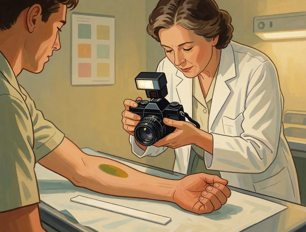
One rule is not a calendar, and the colors were shakier still.
Nobody could agree what they were seeing
Langlois and Gresham could not even score green. Deep blood reads blue, shallow bilirubin reads yellow, and where they overlap your eyes obligingly report green. All their subjects had pale skin, too; on darker skin the colors proved too difficult to make out.
Every bruise on Earth runs the same colors in the same order, and not one of them keeps to a schedule.
By 2005 Maguire and three colleagues had run a systematic review of every published study. Estimating a bruise’s age from its color has no scientific basis, they wrote, and belongs nowhere near a child protection case.
So the sequence is real and the timetable was imaginary. Your bruise will go purple, then green, then yellow, and it will take precisely as long as it takes.
Make a bruise in a glass of water
The chemistry needs a macrophage. The purple only needs a glass.
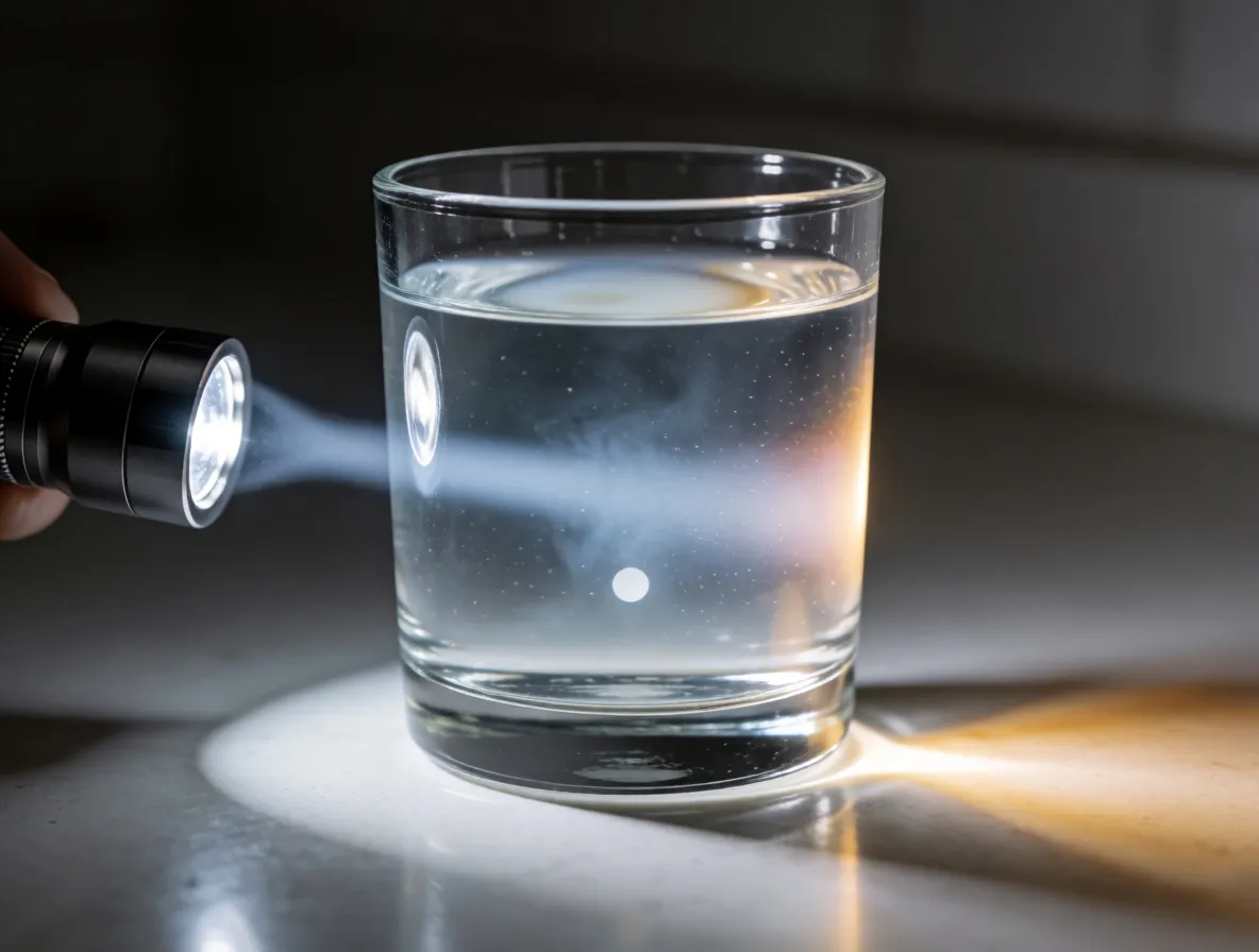
From the side that water glows faintly blue; end on, the flashlight turns orange. Neither color is in the glass. Milk scatters blue sideways more readily than red, so blue leaks out while red carries on, exactly as in your arm.
The yellow never really leaves
Bilirubin does not stay in your leg. It dissolves out, rides the blood to your liver, and is stirred into bile, the yellow-green juice your liver squirts into your intestine to break up fat, and then it leaves you altogether.
Which means the yellow was never something your bruise invented. Every red blood cell wears out after roughly four months, and each is dismantled in precisely this order: the ring, the cut, the green, the yellow. Around two million discs a second, whether you bump into anything or not.
You never see it. That demolition happens inside your spleen, your liver and your bone marrow, where nothing is visible. A bruise is the one occasion when the job is done immediately beneath your skin, where you get to stand and watch.